Stomach histology
The stomach is a key part of the gastrointestinal (GI) tract, sitting between the esophagus and duodenum. Its functions are to mix food with stomach acid and break food down into smaller particles using chemical and mechanical digestion.
The stomach can perform these roles due to the layers of the stomach wall. These are the gastric mucosa, submucosa, muscularis externa and serosa. All parts of the GI tract tend to follow this same pattern of tissue layer arrangement, which means that the stomach is essentially just a widening of the GI tube. These layers are best observed when you’re looking at the microanatomy, or histology, of the stomach.
| Mucosa | Surface mucous cells: simple columnar epithelium Gastric pits: surface mucous cells Gastric glands: parietal, chief, enteroendocrine cells Lamina propria: connective tissue Muscularis mucosa: two smooth muscle layers |
| Submucosa | Connective tissue, submucosal (Meissner’s) plexus |
| Muscularis externa | Smooth muscle layers (longitudinal, circular, oblique), myenteric (Auerbach’s) plexus |
| Serosa | Connective tissue, mesoderm |
| Mnemonic | M.S.M.S |
Histology may not be the easiest to digest, but we will help you sink your gnashers right into this topic and break it down into small logical sections. No hydrochloric acid needed! If you still find it daunting, why not brush up on your histology basics first?
Stomach wall
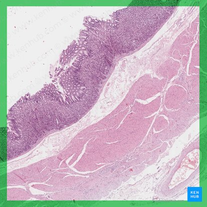
The stomach wall consists of 4 layers of tissue. From deep (external) to superficial (internal) these are the serosa, muscularis externa, submucosa and mucosa. This layered arrangement follows the same general structure in all regions of the stomach, and throughout the entire gastrointestinal tract. The outer layer of the stomach wall is smooth, continuous with the parietal peritoneum. The inner wall (mucosa and submucosa layers) is thrown into folds known as rugae, or gastric folds, which allow the stomach to distend upon the entry of the food. A bolus of food enters the stomach from the esophagus. The various tissue layers of the stomach wall then combine their functions to digest the bolus into a viscous, pulpy fluid called chyme. Chyme is directed into the duodenum of the small intestine for further digestion and absorption.
You can easily remember the four layers of the stomach wall using the mnemonic ‘M.S.M.S‘. It stands for: Mucosa, Submucosa, Muscularis externa & Serosa.
Although the stomach is anatomically divided into four regions, histologically we identify only three; cardia, fundus and pylorus. This is because the fundus and body are histologically identical. Let’s now take a closer look at the 4 layers of the stomach, as well as their regional variations.
Mucosa
The innermost layer of the stomach wall is the gastric mucosa. It is formed by a layer of surface epithelium and an underlying lamina propria and muscularis mucosae. The surface epithelium is a simple columnar epithelium. It lines the inside of the stomach as surface mucous cells and forms numerous tiny invaginations, or gastric pits, which appear as millions of holes all throughout the stomach lining. These gastric pits are important as they are connected to the various glands of the stomach.
There are 3 types of glands found in the stomach; cardiac, gastric and pyloric, named after the region in which they are found. These glands produce the digestive enzymes and mucous secretions of the stomach. The gastric glands of the fundus/body have the important role of producing digestive gastric juice while the cardiac and pyloric glands predominantly produce mucous secretions which protect the stomach from the harsh effects of the digestive acid and prevent stomach self-digestion.
Surface mucous cells
The surface mucous cells, also known as foveolar epithelium, are the simple columnar epithelium lining the lumen of the stomach. They secrete alkaline, highly viscous mucus, which closely adheres to the cellular surface.
The mucus protects the stomach lining by minimising the abrasion from food particles and forming a physical barrier from the hydrochloric acid, in which the mucous cells are constantly bathed. Without these mucous secretions the stomach acid would literally burn holes through the stomach wall! They stain fairly lightly in H&E sections due to the mucin they contain, because it doesn’t pick up either of the stains particularly well.
Gastric pits
Gastric pits are formed by invaginations of the surface epithelium. Gastric pits connect to gastric glands and thus allow the glandular products to be delivered into the stomach lumen. The pits are lined with the same mucus secreting surface epithelium that faces the stomach lumen. In a histological section these will often be cut transversely rather than longitudinally, so will appear as small circular openings, rather than tubular invaginations.
Gastric glands
Gastric glands open into the base of gastric pits. They are found throughout the entire inner surface of the stomach and are divided into 3 types depending on the region in which they are found. Gastric glands proper (principal glands) are found in the fundus/body of the stomach. The cells of these glands produce around two litres of gastric juice a day. The mucus secreting pyloric glands are only associated with the pyloric antrum and cardiac glands are located only within the cardia of the stomach.
Gastric pits and gastric glands are made up of the same 5 cell types: mucous neck cells, stem cells, parietal (oxyntic) cells, chief (zymogenic) cells and enteroendocrine cells. You can see these cells, as well as the substances they secrete, summarised in the table below.
| Mucous neck cells | Mucus secretion (less alkaline than that of the surface epithelial mucous cells) Round nuclei and apical secretory granules Shorter than surface mucous cells |
| Stem cells | Replace damaged cells |
| Parietal (oxyntic) cells | Intrinsic factor production Hydrochloric acid (HCl) secretion Large round or pyramidal cells Highly acidophilic (stain pink) Central rounded nuclei |
| Chief (zymogenic) cells | Pepsinogen and gastric lipase secretion Found in lower regions of gastric glands Basophilic (stain blue) |
| Enteroendocrine cells | Gastrin (released into blood) Single cells (don’t form clusters) |