Small intestine
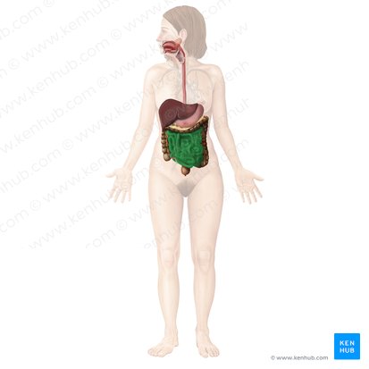
The small intestine is the longest part of the digestive system. It extends from the stomach (pylorus) to the large intestine (cecum) and consists of three parts: duodenum, jejunum and ileum. The main functions of the small intestine are to complete digestion of food and to absorb nutrients.
Dysfunction of the small intestine can bring you some uneasy experiences such as diarrhea while travelling or worse, on a date. This article will discuss the anatomy, function and neurovasculature supply of the small intestines.
| Definition | A part of the alimentary tract which extends from the stomach (pyloric orifice) to the large intestine (ileal orifice) |
| Parts | Duodenum, jejunum, ileum |
| Blood supply | Arteries: celiac trunk, superior mesenteric artery Veins: hepatic portal vein, superior mesenteric vein |
| Innervation | Parasympathetic: vagus nerve (CN X) (through the submucosal (Meissner’s) and myenteric (Auerbach’s) nervous plexuses) Sympathetic: Thoracic splanchnic nerves |
| Function | Final stages of food digestion Absorption of nutrients and water |
| Clinical relations | Diarrhea, obstructive disorders, infectious diseases, neoplastic growths, congenital conditions, duodenal ulcer |
Anatomy
The small intestine is divided into the duodenum, jejunum, and ileum. Together these can extend up to six meters in length. All three parts are covered with the greater omentum anteriorly. The duodenum has both intraperitoneal and retroperitoneal parts, while the jejunum and ileum are entirely intraperitoneal organs. As the small intestine is the main site for the final stages of food digestion and its absorption, its gross and microanatomy are adjusted to that function.
Duodenum
The duodenum by definition is the first part of the small intestine. It extends from the pyloric sphincter of the stomach, wraps around the head of the pancreas in a C-shape and ends at duodenojejunal flexure. This flexure is attached to the posterior abdominal wall by a peritoneal fold called the suspensory muscle (ligament) of duodenum, also called the ligament of Treitz.
The duodenum has four parts: superior (duodenal bulb/ampulla), descending, horizontal and ascending. Among several features of the duodenum, we’ll list the two most important:
- The superior part (duodenal bulb/ampulla) is the only intraperitoneal part, as the hepatoduodenal ligament and greater omentum attach to it.
- The descending part of the duodenum has an opening called the major duodenal papilla (tubercle of Vater). The papilla contains the hepatopancreatic sphincter (sphincter of Oddi, Glissons’ sphincter) which regulates the emptying of the bile from the hepatopancreatic ampulla.
To learn more about the anatomy, histology and function of the duodenum check out our study materials.