2023 Endocrine Images Award Winners
Now in its second year, the competition drew more than 30 entries that were judged by our member volunteers based on aesthetic value and significance to endocrine research. The winning images will be displayed at ENDO 2023, June 15-18 in Chicago, Illinois, and featured through our various print and online media channels.
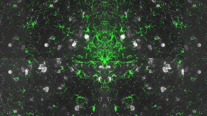
First Place
Anzela Niraula – University of Washington
The image shows microglia (green) and POMC neurons (gray) in close proximity within the arcuate nucleus of the hypothalamus. Microglial regulation of POMC neurons holds significant implications for the pathogenesis of obesity and diabetes.

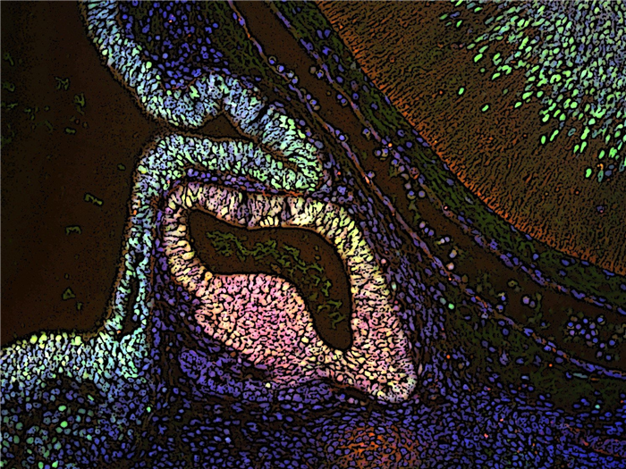
Second Place (tie)
Michelle L. Brinkmeier, Sally Camper, and Deb Gumucio – University of Michigan
The pituitary gland produces hormones that regulate metabolism, fertility, growth, and the stress response. The anterior lobe develops Rathke’s pouch, and the posterior lobe and pituitary stalk develop from overlaying neural ectoderm. Stem cells in both tissues are marked by SOX2 immunostaining (green). The pituitary transcription factor PROP1 (Red) is an important driver of hormone producing cell fate within Rathke’s pouch, where its expression overlaps with SOX2 (yellow). [This is a sagittal slice of an e13.5 mouse embryo.]

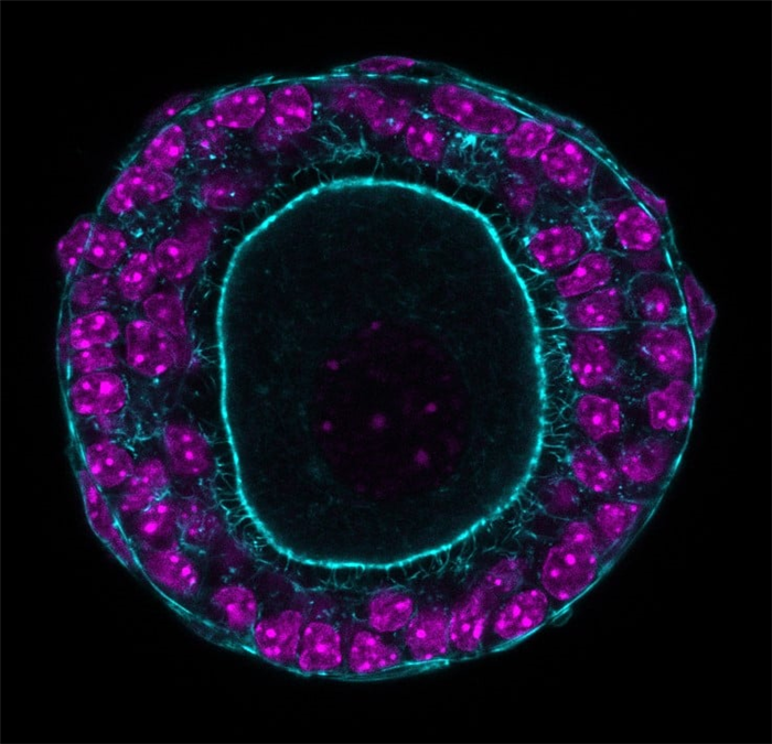
Second Place (tie)
Aubrey Converse – Northwestern University Feinberg School of Medicine
Confocal image of a secondary ovarian follicle (mouse) labeled for DAPI (magenta) and actin (cyan). Ovarian follicles establish actin-rich transzonal projections (TZPs) between the oocyte and supporting steroidogenic granulosa cells. TZPs allow for direct cytoplasmic communication between the oocyte and granulosa cells through gap junctions. Follicle growth and oocyte development are regulated at multiple levels of signaling, including direct cytoplasmic, autocrine, paracrine, and endocrine.

Honorable Mentions
Dima Abdelmannan – Dubai Academic Health Corporation
Image shows a Microscopic specimen of a small nodule from the Thyroid Gland. The Low-power magnification image of the FNA specimen shows abundant watery colloid with low cellularity over a clean background (Papanicolaou stain). This can be also known as “watery colloid”. This type of colloid is present primarily in benign thyroid nodules. The pattern showcases a unique design that resembles a coffee stain on a book page.

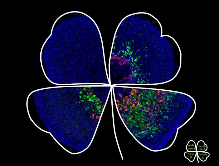
Jeff Huang and Sophia Zheng – Auburn University
The adrenal gland inner cortex expresses thyroid hormone receptor β1 and is responsive to thyroid hormone treatment. In mice, thyroid hormone treatment delays the regression of the adrenal gland’s inner cortex in males and expands the inner cortical domain in females. Cells in the affected area become hypertrophic. Red cells are positive for the X-zone marker 20αHSD. The green signal is CYP2F2. The blue color is DAPI nuclear stain.

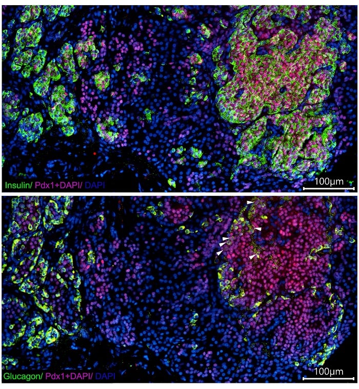
Yuliya Krivova, Alexandra Proshchina, Dmitriy Otlyga, Diliara Gubaeva, and Maria Melikian – Petrovsky National Research Centre of Surgery
The image demonstrates the expression of β-cell specific transcriptional factor Pdx1 in the insulin-positive cells (upper image) and some glucagon-positive cells (bottom image, arrowheads) in the focal lesion of infant with congenital hyperinsulinism.

Natalia Pascuali – Veiga Lab (Department of Pathology – University of Illinois at Chicago)
“A sea of possibilities.” Spatial lipidomics of a human ovary visualized using state-of-the-art mass spectrometry imaging (MSI). The different colors (magenta, blue, and green) illustrate the abundance and localization of three distinct lipid species. We found these lipids to be highly enriched in different ovarian compartments (magenta – theca layer; green – granulosa layer; blue – stroma). Numerous primordial follicles from the ovarian reserve can be observed to the left. To our knowledge, this is the first MSI analysis describing physiological localization of lipids in the human ovary. Therefore, his study could pave the way to finding lipid markers for various ovarian cell types.

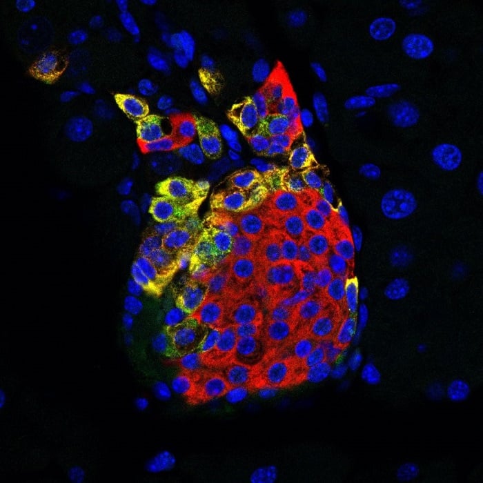
Rachel Reinert – University of Michigan
Whooo is ready to learn about prohormone processing? Pancreatic islets can make fun shapes under the microscope, like this one mimicking a perched horned owl. These islet cells were labeled for glucagon (green) and the processing enzyme prohormone convertase 2 (PC2, red) in addition to the nuclear stain DAPI (blue).

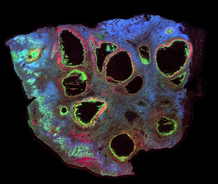
Salimkhanov Rustam, Kim Ekaterina, Urusova Liliya, and Shirshin Evgeny – Endocrinology Research Centre
Confocal image of an adult parathyroid adenoma with intrathyroidal location cryosection showing different intensity and spectral characteristics of autofluorescence of thyroid (upper part) and parathyroid gland (lower part) tissues. Combination of brightfield and epifluorescence imaging.
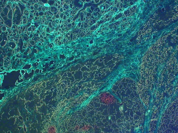

Drayson Jaffee, Anthony Yin, Ian Jaffee – California Pacific Medical Center
Metastatic medullary thyroid carcinoma to the liver: The original image was a 400X hematoxylin and eosin (H&E) stained slide for conventional light microscopy documenting pathology confirmation of metastatic medullary thyroid carcinoma to the liver. The image was subsequently modified with solarizing effect and central ballooning expansion of the neoplastic deposit. Peritumoral endothelialized capillaries are enhanced by aqua staining; and erythrocytes are further delineated by magenta staining, capturing the inherent capacity for these rare malignancies to characteristically disseminate via the peripheral vascular system.