Pituitary gland (hypophysis)
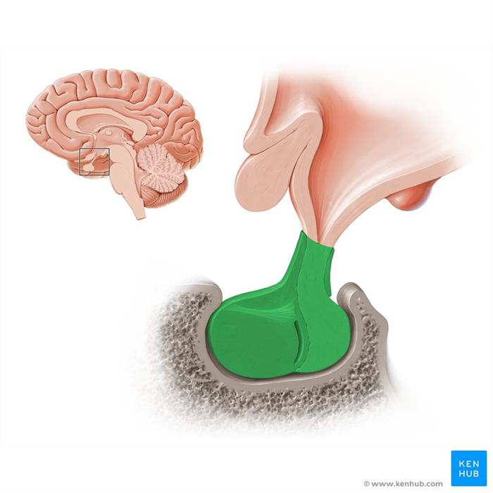
The pituitary gland (hypophysis), is the master gland of the endocrine system. It is an ovoid-shaped structure, located in the sella turcica of sphenoid bone. The pituitary gland is anatomically and functionally closely related to the hypothalamus.
The pituitary gland is made of two active lobes; anterior and posterior.
- The anterior lobe of the pituitary gland, also known as the adenohypophysis, produces and secretes the majority of pituitary hormones. Its function is controlled by the releasing-hormones of the hypothalamus.
- The posterior lobe (neurohypophysis) doesn’t produce any hormones, but it rather releases two hormones that are initially produced in the nuclei of the hypothalamus.
The main function of the pituitary gland is to produce hormones that regulate many vital functions and processes, such as metabolism, growth, sexual maturation, reproduction, blood pressure and many other vital physical functions and processes. The hormones secreted by the gland affect nearly every body system (e.g. other endocrine glands, cardiovascular system, digestive system, reproductive system, etc).
This article will discuss the anatomy and function of the pituitary gland.
| Definition | The “master gland” of the endocrine system which controls the rest of the body glands. |
| Structure | Anterior pituitary (adenohypophysis) Posterior pituitary (neurohypophysis) |
| Hormones | Anterior pituitary: Growth hormone (somatotropin), prolactin, follicle-stimulating hormone (FSH), luteinizing hormone (LH), thyroid-stimulating hormone (TSH), adrenocorticotropic hormone (ACTH) Posterior pituitary: Oxytocin, vasopressin |
| Function | Regulation of metabolism, growth, sexual maturation, reproduction, blood pressure, breastfeeding, immune response and many other vital physical functions and processes |
Structure and location
The pituitary gland is located in the pituitary fossa (sella turcica) of the sphenoid bone. Superiorly, the pituitary gland is covered by the circular diaphragma sellae of dura mater. Anteroinferiorly, the gland faces the sphenoid sinus, anterosuperiorly the optic chiasm and laterally the cavernous sinus.
The pituitary gland is connected to the hypothalamus by the infundibulum (pituitary stalk), which is a process that extends inferiorly from the tuber cinereum of the hypothalamus. The infundibulum not only connects the two glands physically, but it also enables the passage of the hypothalamic hormones to the hypophysis as it is traversed by the hypophyseal portal system and hypothalamohypophyseal tract.
The hypophysis has two major parts, adenohypophysis (anterior part) and neurohypophysis (posterior part). These parts differ in their embryological origin, and thus have different histological appearance and functions.
Hypophyseal portal system
The hypophyseal portal system is a vascular system made of tiny blood vessels that connect the adenohypophysis to the hypothalamus. The capillaries and venules of the portal system are fenestrated which allows the smooth exchange of molecules between the blood vessels and the cells of the anterior pituitary. The epithelial cells of adenohypophysis are arranged in cords between vascular sinusoids where the hormonal exchange between the neural tissue and blood happens.
The hypophyseal-portal system originates from the superior and inferior hypophyseal arteries, which are the branches of the internal carotid artery. The former provides the supply mostly for the anterior pituitary, while the latter supplies the posterior pituitary.
The superior hypophyseal arteries form a primary plexus within the infundibulum and median eminence. This plexus consists of many fenestrated capillaries which rejoin and form the hypophyseal portal veins that travel to the anterior pituitary. The portal veins divide and form another plexus in the anterior pituitary; the secondary plexus. This vascular system is very important as it is the direct connection between the site of releasing of hypothalamic hormones (median eminence) and the cells of the adenohypophysis.
To summarize, the hypophyseal portal system consists of the primary and secondary capillary plexuses (beds) in the pituitary gland, plus the intervening portal veins.
Hypothalamohypophyseal tract
The hypothalamohypophyseal tract is a bundle of axons that connects the hypothalamic nuclei with the neurohypophysis. Its function is to carry the two hypothalamic neurohormones oxytocin and antidiuretic hormone to the neurohypophysis, where they are stored and released upon the body’s needs.
Anterior pituitary (adenohypophysis)
The anterior pituitary (adenohypophysis) is made of three distinctive parts:
- Anterior part (distal or glandular part) is the part with the strongest secretory activity. It is composed of follicles that vary in size, but essentially contain three types of cells. These cells are classified according to their histological staining and include chromophils (acidophilic and basophilic) and chromophobes.
- The acidophilic cells further divide into somatotrophs and lactotrophs. The former produce the growth hormone, while the latter produce prolactin.
- The basophilic cells are divided into gonadotrophs (producing FSH and LH), corticotrophs (ACTH) and thyrotropes (TSH).
- The chromophobes stain weakly, and they are the progenitor cells.
Anterior pituitary hormones
The anterior pituitary secretes five different endocrine hormones, and releases them into the bloodstream. These hormones are the:
Hormones of the anterior pituitary
Somatothrops Hormone: Somatotropine (Growth hormone; GH)
Function: Stimulates growth in epiphyseal plates of long bones via insulin-like growth factors (IGFs) produced in liverLactotrophs (mamotrophs) Hormone: Prolactin
Function: Milk secretion from the mammary glandsThyrothrops Hormone: Thyrotropin (TSH)
Function: Stimulates thyroid hormone synthesis, storage and secretionCorticotrophs Hormones: Adrenal corticotropin (ACTH), lipotropin (LPH)
Function: Stimulates secretion of adrenal cortex hormones (ACTH); Regulates lipid metabolism (LPH)Gonadotrophs Hormones: Follicle stimulating hormone (FSH), luteinizing hormone (LH)
Function: Promotes development of ovarian follicle, estrogen secretion in females, spermatogenesis in males (FSH); Promotes ovarian follicle maturation, progesterone secretion in females, interstitial cell androgen secretion in males (LH)Regulation of the anterior pituitary function
The hypothalamus is the main regulator of the anterior pituitary function. It is one of the very few areas in the brain that is not sealed off from the cerebral bloodstream by the blood–CSF barrier, and therefore can monitor and respond to changes in the body temperature, energy needs, or electrolyte balance. These changes are specifically detected by the hypothalamic nuclei, including the arcuate, paraventricular and ventromedial nuclei and the medial preoptic and paraventricular regions.
The hypothalamus regulates the activity of the anterior pituitary by releasing its stimulating and/or inhibitory hormones that include:
- Corticotropin-releasing hormone (CRH)
- Growth hormone-releasing hormone (GHRH)
- Gonadotropin-releasing hormone (GnRH)
- Thyrotropin-releasing hormone (TRH)
- Dopamine (DA)
- Somatostatin (SS), also known as the growth hormone inhibiting hormone (GHIH)
These hormones are secreted into the bloodstream and sent to the adenohypophysis via the hypophyseal portal system to stimulate or inhibit the secretory activity of its cells. All the hypothalamic “-releasing” hormones have a stimulatory effect, while the “-inhibiting” hormones have inhibitory effects. The secretory activity of the hypothalamus and hypophysis is regulated by the negative feedback mechanism. There are two negative feedback loops that affect the hypothalamic-pituitary axis:
- Long loop feedback: When the blood level of hormones from the peripheral glands reaches the homeostatic/physiological value, those hormones signal to the pituitary and hypothalamus that it’s time to stop the secretion of releasing and stimulating hormones until the values lower again.
- Short-loop feedback: The rise of pituitary hormones blood levels inhibits the synthesis and/or release of the related hypothalamic hormones.