Tissue types

A tissue is a group of cells, in close proximity, organized to perform one or more specific functions.
There are four basic tissue types defined by their morphology and function: epithelial tissue, connective tissue, muscle tissue, and nervous tissue.
- Epithelial tissue creates protective boundaries and is involved in the diffusion of ions and molecules.
- Connective tissue underlies and supports other tissue types.
- Muscle tissue contracts to initiate movement in the body.
- Nervous tissue transmits and integrates information through the central and peripheral nervous systems.
- Epithelial tissue
- Cell surfaces
- Tissue structure
- Specialized epithelial tissue
- Connective tissue cells
- Connective tissue fibers
- Connective tissue classification
- Skeletal muscle
- Cardiac muscle
- Smooth muscle
- Neurons
- Tissue
- Epithelial tissue
- Connective tissue
- Muscle tissue
- Nervous tissue
Epithelial tissue
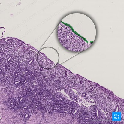
Epithelial tissue is a highly cellular tissue that overlies body surfaces, lines cavities, and forms glands. In addition, specialized epithelial cells function as receptors for special senses (smell, taste, hearing, and vision). Epithelial cells are numerous, exist in close apposition to each other, and form specialized junctions to create a barrier between connective tissues and free surfaces. Free surfaces of the body include the outer surface of internal organs, lining of body cavities, exterior surface of the body, tubes and ducts. The extracellular matrix of epithelial tissue is minimal and lacks additional structures. Although epithelial tissue is avascular, it is innervated.
Test what you already know about epithelial tissue with our quiz!
Cell surfaces
The cells of epithelial tissue have three types of surfaces differentiated by their location and functional specializations: basal, apical, and lateral.
Basal surface
The basal surface is nearest to the basement membrane. The basement membrane itself creates a thin barrier between connective tissues and the most basal layer of epithelial cells. Specialized junctions called hemidesmosomes secure the epithelial cells on the basement membrane.
Apical surface
The apical surface of an epithelial cell is nearest to the lumen or free space. Apical cell surfaces may display specialized extensions. Microvilli are small processes projecting from the apical surface to increase surface area. They are heavily involved in diffusion in the proximal convoluted tubule of the nephron and in the lumen of the small intestines.
Cilia are small processes found in the respiratory tract and female reproductive tract. Their complex structure facilitates movement that brushes small structures through the lumen of either the trachea or Fallopian tubes. Stereocilia are similar to cilia in size and shape, however they are immotile and more frequently found in the epithelium of the male reproductive tract, specifically in the ductus deferens and the epididymis.
Lateral surfaces
The lateral surfaces of epithelial cells are located between adjacent cells. The most notable lateral surface structures are junctions. Adhering junctions link the cytoskeleton of neighboring cells to produce strength in the tissue. Desmosomes can be thought of as spot-welding for epithelial tissues. They are usually located deep to adhering junctions and are found in locations subject to stresses. For example in the stratified epithelium of the skin.
Tight junctions form a solid barrier to prevent movement of molecules between adjacent epithelial cells. Tight junctions are found in the simple columnar epithelium of the gut tube to regulate absorption of nutrients. Finally, gap junctions perform the opposite function. Gap junctions allow small molecules and structures to pass freely between cells. For example, gap junctions in cardiac muscle tissue allow for coordinated contraction of the heart.
Summary of epithelial tissue surfaces and characteristics
Characteristics Highly cellular, function as receptors, form a barrier, minimal extracellular matrix, avascular, innervated, Basal surface Basement membrane, hemidesmosomes Apical surface Microvilli, cilia, stereocilia Lateral surface Adhering junctions, desmosomes, tight junctions, gap junctions Tissue structure
Two major characteristics of epithelial tissue divide it into subclasses: the shape of the cells and the presence of layers.
- Squamous – cells are flattened, can be keratinized or nonkeratinized, involved in protection and diffusion, found in capillary walls and skin
- Cuboidal – cells are cube-shaped, can be found forming tubes in the nephrons of the kidney, involved in secretion and absorption
- Columnar – cells are rectangular, cilia are often present, involved in absorption, secretion, protection, and lubrication, form the inner lining of the gut tube
- Simple – one layer of cells
- Stratified – two or more layers of cells
- Pseudostratified – simple epithelia that appear to be stratified when viewed in cross-section though they are only one layer of cells
Specialized epithelial tissue
- Transitional epithelium – distends tissues of urinary tract
- Keratinized stratified squamous epithelium – makes up the epidermis of skin
- Nonkeratinized stratified squamous epithelium – found in regions subject to abrasion, for example oral mucosa and vaginal lining
- Pseudostratified ciliated columnar epithelium – lines the inner surface of the trachea
- Endothelium – lines the inner surface of blood vessels
- Ependymal cells – present in the nervous system
Connective tissue
Connective tissue is the most abundant tissue type in the body. In general, connective tissue consists of cells and an extracellular matrix. The extracellular matrix is made up of a ground substance and protein fibers. So, in a more detailed way, all connective tissue apart from blood and lymph consists of three main components: cells, ground substance and fibers.
Summary of connective tissue
Cell types Structural, immunological, defense, energy reservoir Fibers Collagen, reticular, elastic Classification Proper: Loose; dense (regular, irregular) connective tissue
Embryonic: Mesenchymal; mucous connective tissue
Specialized: Cartilage; adipose; bone; bloodConnective tissue cells
The cells originate from mesenchyme, a loosely organized embryonic tissue featuring elongated cells in a viscous ground substance. Connective tissue cells do not oppose each other but rather are separated by a large extracellular matrix.
- Structural – fibroblasts, chondroblasts, osteoblasts, odontoblasts
- Immunological – plasma cell, leukocytes, eosinophils
- Defense – neutrophils, mast cells, basophils, macrophages
- Energy reservoir – adipose cells
Connective tissue fibers
The ground substance of connective tissue contains structural proteins called fibers. There are three types of connective tissue fibers:
- Collagen fibers are the most abundant fiber type. They have a high tensile strength but are also flexible. Collagen fibers are made up of many subunits, called collagen fibrils, that appear striated under electron microscopy. There are many types of collagen and the collagen types present in a tissue give it unique characteristics. For example, type I collagen provides resistance to stretch in bone tissue, while type IV collagen makes up the suprastructure of the basement membrane.
- Reticular fibers are thinner than collagen fibers. They are found in extensive networks and provide structural support and framework. Reticular fibers do not stain with regular H&E stain and a silver stain is needed to stain fibers black, making them visible.
- Elastic fibers are also thinner than collagen. They are strong but can be stretched up to 150% of their original length without breaking. When tension is released they are able to return to their original shape. Elastic fibers are found in skin, blood vessels and lung tissue.
Connective tissue classification
Classification of connective tissue is based upon two characteristics: the composition of its cellular and extracellular components and its function in the body. Tissues are either classified as proper, embryonic, or specialized.
Proper connective tissues
Proper connective tissues include loose connective tissue, often referred to as areolar tissue, and dense connective tissue. Loose connective tissue consists of thin, loosely arranged collagen fibers in a viscous ground substance.
Dense connective tissue can be further classified into dense regular connective tissue and dense irregular connective tissue. Dense regular connective tissue makes up tendons and ligaments. Fibers are densely packed and organized in parallel to create a strong tissue capable of withstanding the pull of muscle and bone in movement. Dense irregular connective tissue also contains abundant fibers but lacks the directionality of dense regular connective tissue fibers. The high number of fibers provides strength however the disorganized pattern of fibers allows for flexibility. Dense irregular tissue is associated the hollow organs of the digestive tract.
Do you need help identifying tissues? Try our tissue quizzes and worksheets to simplify your learning, cement your knowledge and ace your histology exams!
Embryonic connective tissue
Embryonic connective tissue, derived from mesoderm, is the precursor to many connective tissues in the adult body. It is categorized into two subtypes: mesenchyme and mucous connective tissue. Mesenchyme is found within the embryo. Mesenchymal cells are spindle shaped with processes extending from either end. The cell processes connect to those of other mesenchymal cells through gap junctions. Very thin, scattered collagen fibers are present, but they are not particularly strong reflecting the limited stress placed on the tissues of the developing embryo.
Mucous connective tissue is found in the umbilical cord. The cells of mucous connective tissue are spindle shaped and relatively sparse. A nearly gelatinized ground substance called Wharton’s jelly makes up most of the extracellular matrix between the cells and collagen fibers.
Do you want to find out more about connective tissue in a more visual way? Follow along with the following study units:
Specialized connective tissues
Cartilage, adipose tissue, bone, and blood are specialized connective tissues. Adipose cells, or adipocytes, are specialized cells that store fat and synthesize hormones, growth factors, and some inflammatory mediators. They are located in loose connective tissue either as individual cells or in clusters. When adipocytes are clustered in large numbers they are referred to as adipose tissue.
Bone tissue is unique in that its extracellular matrix is mineralized. Calcium phosphate, in the form of hydroxyapatite crystals, is responsible for the mineralization of bone and creates a very strong tissue able to support and protect the body.Blood is a fluid connective tissue that transports gases, nutrients, and wastes throughout the body. The fluid extracellular matrix of blood is made up of plasma, which constitutes slightly more than half of the tissue volume. The cells of blood tissue are classified as erythrocytes, leukocytes, and thrombocytes. Erythrocytes, or red blood cells, carry oxygen and carbon dioxide through the cardiovascular system. Leukocytes, or white blood cells, are responsible for the immune and allergic responses. Thrombocytes, or platelets, form clots and initiate the repair of injured blood vessels.
Details about specialized connective tissues are provided below:
Muscle tissue
Muscle tissue is both extensible and elastic, in other words, it can be stretched and returned to its original size and shape. The cells of muscle tissue are unique in that they are contractile, or capable of contraction. This contraction is a result of sliding actin and myosin filaments. Muscle tissue is easily distinguishable by its highly organized bundles of cells. Although there are three types of muscle tissue with unique cell morphologies, the fiber bundles of each tissue type are arranged in parallel oriented on the long axis and are distinct from surrounding connective tissue. Muscle is classified according to the appearance of the contractile cells.
Summary of muscle tissue
Characteristics Extensible, elastic, contractile, organized into bundles Skeletal Rapid and strong contraction; large, cylindrical, elongated cells; syncytium; peripheral and ovoid nuclei; striated; present in voluntary skeletal muscles Cardiac Strong contraction; striated; single and centrally located nucleus, connected by gap junctions and intercalated discs; syncytium; found in the myocardium Smooth Weak and slow contractions; spindle shaped cells; single and central nucleus; nonstriated; found in involuntary muscles (viscera) The three types of muscle tissue are: skeletal muscle, cardiac muscle, and smooth muscle tissue.
Skeletal muscle
Skeletal muscle is responsible for the voluntary movement of the body. For example, movement of the limbs, skin of the face, and orbits. Contraction of skeletal muscle tissue is rapid and strong. Cells are large, cylindrical, and elongated. In embryonic development, myoblasts fuse together to form one larger muscle cell, resulting in syncytial, multinucleated cells. Nuclei of skeletal muscle cells are peripheral and ovoid. When viewed under a microscope, the arrangement of actin and myosin gives skeletal muscle a striated appearance.
Cardiac muscle
Cardiac muscle is found in the heart wall also known as myocardium. Like skeletal muscle, actin and myosin also give cardiac muscle a striated appearance. The movement that cardiac muscle cells provide is involuntary and coordinated by gap junctions. A major defining characteristic of cardiac muscle tissue is the presence of intercalated disks. Cardiac muscle cells are elongated and branched. Intercalated disks are present at the junctions between two cells. Although gap junctions allow this tissue to function as a syncytium, each cell has one, centrally located nucleus.
Smooth muscle
Smooth muscle tissue is associated with arteries and tubular organs such as the intestinal tract. This type of tissue provides weak, slow involuntary movements. Smooth muscle cells are spindle shaped with one central nucleus. The contractile fibers of smooth muscle cells are arranged perpendicular to each other rather than in parallel, therefore smooth muscle tissue does not appear striated.
Master the histology of muscle tissue with the following resources:
Nervous tissue
Neurons
Cells of the nervous system are highly specialized to transmit electrical impulses around the body. There are two main types of cells found in nervous tissue: neurons and glia.
Neurons tend to have a large cell body, or soma, and long projections used in transmitting information. These projections are referred to as axons or dendrites. Axons send impulses away from the soma and dendrites carry incoming information. Neurons are most easily identified by their axons in either longitudinal or cross-sectional slide. Groups of neurons are referred to as ganglia in the peripheral nervous system and as nuclei in the central nervous system.
Summary of nervous tissue
Neurons Function: transmission of electrical impulses
Structure: soma (cell body), axons (transmit impulses away from soma), dendrites (transmit incoming impulses)
Organization: ganglia (PNS) and nuclei (CNS)Glia Function: support and nourish neurons
Astrocytes: support synapses, form a protective barrier around blood vessels
Oligodendrocytes: insulate axons and increase impulse projection in the CNS
Schwann cells: oligodendrocytes equivalents in the PNS
Microglia: defend the nervous systemGlia are the supporting cells of nervous tissue and significantly outnumber neurons. These cells differ by region of the nervous system. Astrocytes support neurons, especially near synapses, and provide a protective barrier surrounding blood vessels. Oligodendrocytes are found in the white matter of the central nervous system. Large projections from these cells wrap around the axon of a neuron insulating it to allow for faster projection of impulses.
In the peripheral nervous system, Schwann cells accomplish the same task. Oligodendrocytes and Schwann cells are useful in identifying nervous tissue because the sheathing they provide appears as a thick layer surrounding a tubular axon. Microglia are the macrophages of the nervous system. These cells constantly survey nervous tissue to destroy invaders and clear cell debris.
Nervous tissue exhibits a fluid-filled extracellular space through which ions and neuromediators travel to transmit impulses. Because the generation of action potentials requires a specific concentration of ions, the extracellular environment is highly regulated by glia. Capillaries passing through nervous tissue are completely surrounded by glia to form the blood brain barrier.
Are you curious to find out more about the nervous tissue? You have come to the right place!
Highlights
Tissue
A tissue is a group of cells, in close proximity, organized to perform one or more specific functions. There are four basic tissue types defined by their morphology and function:
Epithelial tissue
Epithelial tissue is a highly cellular tissue that overlies body surfaces, lines cavities, and forms glands. It is avascular but innervated. Epithelial cells exist in close apposition, forming a barrier between connective tissues and free surfaces. Their surfaces face basally, apically and laterally, with each having distinctive features. Specialized epithelial tissue also exists.
Connective tissue
Connective tissue is the most abundant tissue type in the body. It consists of cells, that originate from mesenchyme, and an extracellular matrix. The extracellular matrix is made up of a ground substance and protein fibers. There are several important cell types and three main fibers: collagen, reticular and elastic. Classification of connective tissue into three broad types is based upon the composition of its cellular and extracellular components and its function in the body.
Muscle tissue
Muscle tissue is both extensible and elastic. The cells are contractile and are highly organized into fiber bundles. Muscle is classified according to the appearance of the contractile cells, into three types: skeletal, cardiac and smooth. The first two types have a striated appearance due to the parallel orientation of the fiber bundles.
Nervous tissue
Cells of the nervous system are highly specialized to transmit electrical impulses around the body. There are two main types: neurons and glia. Neurons tend to have a large cell body and projections carrying information to (dendrites) and from (axons) the cell body itself. Groups of neurons are referred to as ganglia (PNS) and nuclei (CNS). Glia are the supporting cells of nervous tissue. They consist of astrocytes, oligodendrocytes/Schwann cells and microglia.
Sources
All content published on Kenhub is reviewed by medical and anatomy experts. The information we provide is grounded on academic literature and peer-reviewed research. Kenhub does not provide medical advice. You can learn more about our content creation and review standards by reading our content quality guidelines.
- M. H. Ross: Histology: A Text and Atlas, 6th edition, Lippincott Williams & Wilkins (2011), p. 98-101; 159-172
- S. G. Waxman: Clinical Neuroanatomy, 27th edition, McGraw-Hill Education (2013), p. 7-14
- Epithelia Lab: Histology at Yale
Tissue types: want to learn more about it?
Our engaging videos, interactive quizzes, in-depth articles and HD atlas are here to get you top results faster.
“I would honestly say that Kenhub cut my study time in half.” – Read more. Kim Bengochea, Regis University, Denver
© Unless stated otherwise, all content, including illustrations are exclusive property of Kenhub GmbH, and are protected by German and international copyright laws. All rights reserved.
Register now and grab your free ultimate anatomy study guide!