Pituitary Gland Anatomy and Basics
The pituitary gland sits at the base of the brain and is responsible for secreting hormones that regulate various body functions. Tumors of the pituitary gland cause symptoms because of dysregulation of the hormones secreted by the pituitary gland or from pushing on important structures in the brain such as the eye (optic) nerves.
Herein, we will discuss what the pituitary gland is, where it is located, and how these characteristics impact the symptoms that may develop because of pituitary tumors.
Where Is the Pituitary Gland Located?
The pituitary gland is located at the base of the brain and rests in a bony structure within the skull called the sella turcica. This formation of bone surrounds and protects the pituitary gland. When looking in the mirror, you can approximate the location of the pituitary gland directly behind the midpoint between your eyes.
The pituitary gland has two distinct sections, the anterior and posterior pituitary. The anterior pituitary gland is sometimes referred to as the adenohypophysis while the posterior pituitary gland is referred to as the neurohypophysis. These alternative names are related to the type of tissue each portion is made of.
Together, the pituitary gland is often called the master gland. This is because the pituitary gland controls the release of most of the hormones in the body. Detailed information about pituitary hormones and function are discussed in another article.
What Is Near the Pituitary Gland?
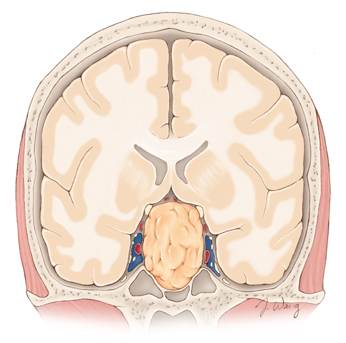
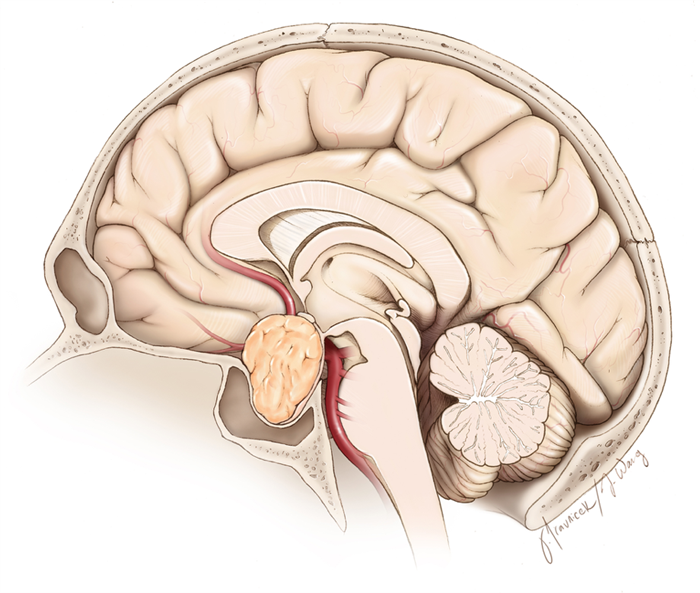
Figure 1. A pituitary tumor (orange) is demonstrated in coronal (front view) and sagittal (side view) planes of the head. A thin rim of the normal gland is usually draped over the tumor.
The pituitary gland is surrounded on either side by the cavernous sinus (blue structures in Figure 1). The cavernous sinus is a small, hollow space within the base of the skull that is filled with venous blood through which important nerves and blood vessels travel. The blood vessels that pass through the cavernous sinus include a section of the carotid artery as it enters your skull to provide blood and oxygen to your brain.
In addition to the carotid artery, the nerves that control the movement of your eyes and sensation to your face pass through the cavernous sinus. The nerves to the eyes for vision are draped over the top of the gland. The proximity of the pituitary gland to these nerves and other structures explains the symptoms someone may experience with a pituitary tumor.
In general, pituitary tumors cause symptoms by either overproducing usual pituitary hormones or not producing any symptomatic hormones but enlarging to the point of compressing the surrounding normal structures.
If the tumor is causing overproduction of hormones, then lactation (unusual breast secretions), obesity, enlargement of hands/fingers or other hormonal abnormalities can occur.
One of the most common symptoms that patients experience because of pituitary tumors is a change in vision, often described as a loss of peripheral vision. This is because the optic chiasm travels directly above the pituitary gland. The optic chiasm is a tract of nerves that connects your eyes to your brain, allowing you to see. Compression of this optic chiasm by a tumor may interrupt the communication between the eyes and brain, resulting in visual changes.
How Can You Tell What Is Normal Pituitary Gland and What Is Tumor?
Evaluation of a pituitary tumor commonly includes a magnetic resonance imaging (MRI) study to visualize the tumor around the pituitary gland. This study will likely include the use of contrast. Contrast is a type of dye that helps differentiate the normal gland from pituitary tumor. As dye is injected into the bloodstream, the normal tissue of the pituitary gland and that of tumor appear differently.
Other findings on imaging that help differentiate normal gland from tumor include the shape of the gland. While normal pituitary gland is typically smooth in appearance, a tumor may cause the edges of the pituitary gland to become distorted or bumpy.
You may hear a pituitary tumor referred to as a microadenoma or a macroadenoma. This is related to the size of the tumor as demonstrated on imaging. Microadenomas are less than 10 mm in diameter while macroadenomas are greater than 10 mm in diameter. For reference, a normal pituitary gland has a diameter of approximately 10 mm, about the size of a kidney bean.
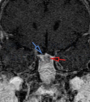
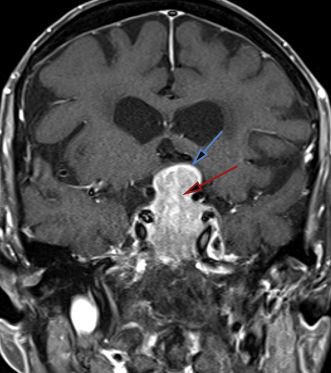
Figure 2. The top image demonstrates a coronal MRI with contrast. The pituitary microadenoma (red arrow) is pushing the compressed rim of normal gland up (blue arrow.) The bottom image shows a coronal MRI with contrast of a pituitary macroadenoma; the tumor (red arrow) is pushing the rim of pituitary gland up as well. Note the difference in the size of microadenomas and macroadenomas.
What Is Transsphenoidal Surgery?
Transnasal transsphenoidal surgery is the common surgical technique for the removal of pituitary tumors. This technique is called transnasal, transsphenoidal because it requires the surgeon to pass through the nose and then the sphenoid sinus to reach the pituitary gland. This approach is commonly used because it allows the most direct route to the pituitary gland that rests behind the nose.
The sphenoid sinus, located behind the nose, is a hollow area that produces mucus to keep your nose from becoming dry. In transnasal, transsphenoidal surgery the surgeon will pass a camera and surgical instruments through the nostrils to the back of the nose. They will then make a small hole in the front portion of the sphenoid sinus to gain access to this hollow chamber.
Once this is done, a small hole is made in the bony, posterior portion of the sphenoid sinus which makes up the floor of the sella turcica. As mentioned previously, the sella turcica is the bony pocket that the pituitary gland rests in. By making a hole in the bottom of the sella turcica, the pituitary gland and its associated tumor may be seen. More detailed information about modern-day treatments of pituitary tumors can be found here.
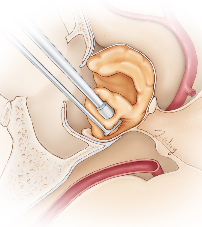
Figure 3. In this side view or sagittal illustration of the surgical corridor, the pituitary tumor (orange) is removed via surgical instruments that are passed through the nose. This procedure is called transnasal transsphenoidal surgery and is very effective in removal of such tumors.
Key Takeaways
- The pituitary gland is located beneath the brain directly behind your eyes in a bony covering called the sella turcica.
- Symptoms of a pituitary tumor occur because of the disruptions or overproduction of the pituitary hormones or from compressing the nearby eye nerves.
- Surgical treatment of pituitary tumors is effective through the transnasal transsphenoidal approach where surgical instruments are passed through the nose to gain access to the pituitary gland/tumor.
Resources
7,000+ complex surgeries over 16 years. Our team has unparalleled experience to provide expert opinions.

Copyright © 2023 Aaron Cohen-Gadol. All Rights Reserved. | Terms of Use | Privacy Policy | Report a Problem