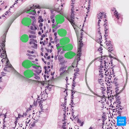
Goblet cells

Goblet cells are unicellular intraepithelial mucin-secreting glands scattered within simple epithelia, such as cuboidal, columnar, and pseudostratified epithelia. Their name corresponds to their shape, as they resemble a goblet, with their narrow bases and wide apex.
Their role is to protect the surface of epithelium, lubricate it, and catch harmful particles. Although protective, goblet cells may be involved in pathophysiology of certain respiratory diseases, such as chronic bronchitis.
This article will discuss goblet cells histology and function.
| Definition | Modified epithelial cells that secrete mucus on the surface of mucous membranes of intestines and airways |
| Location | Respiratory epithelium Intestinal epithelium |
| Staining | Periodic acid Schiff method (PAS) |
| Morphology | Basal part – nucleus, mitochondria, rough endoplasmic reticulum, Golgi complex Apical part – vesicles with mucins |
| Function | Protection and lubrication of epithelial surfaces of the respiratory and digestive tracts by producing mucus |
| Clinical relations | Chronic bronchitis, asthma |
Definition
Goblet cells are modified epithelial cells that secrete mucus on the surface of mucous membranes of organs, particularly those of the lower digestive tract and airways.
Histologically, they are mucous merocrine exocrine glands. What does that mean?
- Goblet cells secrete mucus – mucous glands
- Their product is packed in vesicles inside the cell, and released by exocytosis – merocrine glands
- They release their product on the surface of epithelium rather than in blood – exocrine glands
Location and morphology
Goblet cells are mostly found scattered in the epithelia of the small intestines and respiratory tract. The morphology of goblet cells reflects their function, with the cell containing all the organelles necessary for the production of glycosylated proteins called mucins.
In the respiratory tract, mucins play an important role in catching large particles inhaled by air. Going down the respiratory tree, the number of goblet cells decrease, while the number of club cells increase. This is due to terminal bronchiole needing different kinds of protection which club cells can bring them–breaking down of inhaled toxins that couldn’t be stopped by the mucus of goblet cells.
Struggling with identifying histology slides? Try our histology slide quizzes to cut your studying time!
Besides containing the nucleus and mitochondria, the cytoplasm is especially rich in rough endoplasmic reticulum (RER) and Golgi complexes. Mucins are produced by mucinogen granules in RER and packed into vesicles in Golgi complexes. To make the entire process of synthesis more efficient, goblet cells are polarized, meaning that the organelles have a specific layout within the cytoplasm: the nucleus, mitochondria, RER, and Golgi apparatus are found in the basal portion of the cell; while the vesicles with mucins are located apically, in order to be close to the apical membrane through which their exocytosis occurs.
After exocytosis of the vesicles onto the surface of mucous membranes, the mucins become hydrated and form mucus. Mucus is easily washed out during histological preparation, which is why it stains very poorly with hematoxylin and eosin. But since mucins are heavily glycosylated, meaning they contain a lot of oligosaccharides, they stain purple with the periodic acid-Schiff method (PAS).
Intraepithelial glands study starter pack is waiting for you here.
Functions
In the small and large intestines, goblet cells are dispersed between enterocytes. Their main function here is to produce mucus which protects and lubricates the surface of the intestines. In the respiratory tract, besides protecting the epithelial surface, mucus traps harming particles inhaled with air to protect the airway.
Goblet cells produce small amounts of mucus continually, which is known as basal (constitutive) secretion. Mucus production significantly increases when the epithelium is irritated, for example, by inhaling smoke. This kind of triggered secretion is called stimulated secretion.
Test your knowledge on goblet cells here:
Clinical relations
Chronic bronchitis
Long term smokers often suffer from chronic bronchitis. Since their respiratory tract is constantly irritated by smoke, the number of goblet cells within the respiratory epithelium significantly increases. Thus, mucus production becomes too large to be properly removed by ciliated cells, so it precipitates on the surface of respiratory tract and obstructs the airways. Therefore, patients with chronic bronchitis cough a lot, as their body tries to expel this excess mucus.
Asthma
Increased secretion of mucus is also a pathognomonic feature of asthma–a chronic inflammatory disorder of the respiratory system. There are two categories of asthma depending on the cause: atopic (allergic reaction) and non-atopic (cause is usually unknown). In either type, T-lymphocytes secrete inflammatory substances called cytokines, which stimulate vasodilation, bronchoconstriction, and increased mucus secretion. This increased mucus secretion together with bronchoconstriction significantly narrows the airways and symptomatically presents as a cough, shortness of breath, chest pain and abnormal respiratory sounds (wheezing).
Sources
All content published on Kenhub is reviewed by medical and anatomy experts. The information we provide is grounded on academic literature and peer-reviewed research. Kenhub does not provide medical advice. You can learn more about our content creation and review standards by reading our content quality guidelines.
- Mescher A. l. (2013). Junqueira’s Basic Histology (13th ed.). New York, NY: McGraw-Hill Education
- Ross, M. J., Pawlina, W. (2011). Histology (6th ed.). Philadelphia, PA: Lippincott Williams & Wilkins.
- Kumar, V., Abbas, A. K., Aster, J. C. (2013). Robbins Basic Pathology (9th ed.). Philadelphia, PA: Elsevier Saunders.
Goblet cells: want to learn more about it?
Our engaging videos, interactive quizzes, in-depth articles and HD atlas are here to get you top results faster.
“I would honestly say that Kenhub cut my study time in half.” – Read more. Kim Bengochea, Regis University, Denver
© Unless stated otherwise, all content, including illustrations are exclusive property of Kenhub GmbH, and are protected by German and international copyright laws. All rights reserved.
Register now and grab your free ultimate anatomy study guide!